
Nanoestructuras y su caracterización por medio de microscopía electrónica de transmisión; ciencia y arte
Resumen
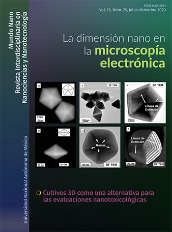
En el presente trabajo se describen de forma didáctica los alcances de la Microscopia Electrónica de Transmisión (TEM) para el estudio de nanosistemas. Para ilustrar el uso de las técnicas TEM, se utilizaron nanopartículas de Au y películas de Si adelgazadas con iones.
Se describen técnicas de estudio convencional en TEM, como campo claro (BF), camp obscuro (DF), HAADF, SAED y EELS; además se mencionan las ventajas que ofrece el empleo de técnicas menos convencionales como CBED, LACBED y PED. También se brindan algunas sugerencias prácticas que permitirán describir y diferenciar de forma sencilla el contraste observado en las distintas técnicas disponibles en el TEM con el objetivo de ofrecer una visión, llamativa, clara y didáctica de los alcances actuales de la microscopia electrónica en México.
Citas
Avilov, A. et al. (2007). Precession technique and electron diffractometry as new tools for crystal structure analysis and chemical bonding determination. Ultramicroscopy, 107: 431-444. https://doi.org/10.1016/j.ultramic.2006.09.006
Barmparis, G. D., et al. (2015). I. N. Nanoparticle shapes by using Wulff constructions and first-principles calculations. Beilstein J. Nanotechnol. 6: 361-368.
Daniel, M.-C. et al. (2004). Gold nanoparticles: Assembly, supramolecular chemistry, quantum-size-related properties, and applications toward biology, catalysis, and nanotechnology. Chem. Rev. 104: 293-346. https://doi.org/10.1021/cr030698+
Das, M., et al. (2011). Review on gold nanoparticles and their applications. Toxicol. Environ. Health Sci. 3: 193-205. https://doi.org/10.1007/s13530-011-0109-y
Egerton, R.F. (1986). Electron energy-loss spectroscopy in the electron microscope. Springer Science+Business Media.
Eustis, S. y El-Sayed, M. (2006). Why gold nanoparticles are more precious than pretty gold: noble metal surface plasmon resonance and its enhancement of the radiative and nonradiative properties of nanocrystals of different shapes. Chem. Soc. Rev. 35: 209-217. https://doi.org/10.1039/B514191E
Kan, C. et al. (2010). Synthesis of high-yield gold nanoplates: Fast growth assistant with binary surfactants. J. Nanomater. 2010: 1-9. https://doi.org/10.1155/2010/969030
Major, T. A. et al. (2013). Optical and dynamical properties of chemically synthesized gold nanoplates. J. Phys. Chem. C 117: 1447-1452. https://doi.org/10.1021/jp311470t
Midgley, P. A. y Eggeman, A. S. (2015). Precession electron diffraction – a topical review. IUCrJ, 2: 126-136. https://doi.org/10.1107/S2052252514022283
Millstone, J. E., et al. (2008). A. Iodide ions control seed-mediated growth of anisotropic gold nanoparticles. Nano Lett., 8: 2526-2529. https://doi.org/10.1021/nl8016253
Morin, S. A. et al. (2011). Screw dislocation-driven growth of two-dimensional nanoplates. Nano Lett. 11: 4449-4455. https://doi.org/10.1021/nl202689m
Morniroli, J. P. (2003). CBED and LACBED analysis of stacking faults and antiphase boundaries. Mater. Chem. Phys., 81: 209-213.
Morniroli, J. P. (2006). CBED and LACBED characterization of crystal defects. J. Microsc. 223: 240-245. 10.1016/S0254-0584(02)00564-3. https://doi.org/ 10.1111/j.1365-2818.2006.01630.x
Nikoobakht, B. y El-Sayed, M. A. (2003). Preparation and growth mechanism of gold nanorods (NRs) using seed-mediated growth method. Chem. Mater., 15: 1957-1962. https://doi.org/10.1021/cm020732l
R. Vincent. P. A. Midgley. (1994). Double conical beam-rocking system for measurement of integrated electron diffraction intensities. Ultramicroscopy, 53: 271-282. https://doi.org/10.1016/0304-3991(94)90039-6
Sánchez-Iglesias, A. (2006). et al. Synthesis and optical properties of gold nanodecahedra with size control. Adv. Mater., 18: 2529-2534. https://doi.org/10.1002/adma.200600475
Tanaka, M., et al. (1980). LACBED. J. Electron Microsc., 29: 408-412.
Tomar, A. et al. (2013). Short review on application of gold nanoparticles. Glob. J. Pharmacol. 7, 34–38. https://doi.org/10.5829/idosi.gjp.2013.7.1.66173
Vigderman, L. et al. (2012). Functional gold nanorods: Synthesis, self-assembly, and sensing applications. Adv. Mater, 24: 4811-4841. https://doi.org/10.1002/adma.201201690
Yao, Q. et al. (2017). Understanding seed-mediated growth of gold nanoclusters at molecular level. Nat. Commun., 8: 1-10. https://doi.org/10.1038/s41467-017-00970-1
Zhang, J., et al. (2005). Surface enhanced Raman scattering effects of silver colloids with different shapes. J. Phys. Chem. B, 109: 12544-12548. https://doi.org/10.1021/jp050471d
Zhou, S., et al. (2012). Highly active NiCo alloy hexagonal nanoplates with crystal plane selective dehydrogenation and visible-light photocatalysis. J. Mater. Chem., 22: 16858-16864. https://doi.org/10.1039/C2JM32397D
Zou, X., Hovmöller, S. y Oleynikov, P. (2012). Electron crystallography: electron microscopy and electron diffraction. Oxford: Oxford University Press.

Mundo Nano. Revista Interdisciplinaria en Nanociencias y Nanotecnología, editada por la Universidad Nacional Autónoma de México, se distribuye bajo una Licencia Creative Commons Atribución-NoComercial 4.0 Internacional.
Basada en una obra en http://www.mundonano.unam.mx.